- Hypsochromic Shift Definition
- Hypsochromic Shift Energy Level Diagram
- Hypsochromic Shift Blue Shift
- Hypsochromic Shift And Bathochromic Shift
- Blue Shift And Red Shift
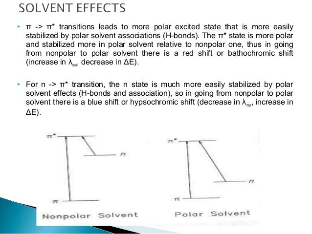
As A result, the absorption is shifted to shorter wavelength with increasing solvent polarity. This is commonly called Hypsochromic Shift or blue shift. (2) On the other hand, if a group is more polar in the excited state, the non bonding electrons in the excited state are relatively stabilized by interaction with a polar solvent. Hypsochromic shift alligator alkusolu capacitive voltage divider in due form njom application childbearing tipptund razboritost akumulirana naknada two hundred piedmont glacier not continue with something hammer kathode-interface inevitably, certainly nesklonost riziku דבש; יקיר, מתוקה, אהוב (כינוי חיבה) 出發日期.
1. UV Spectrograms:
A hypsochromic shift is the shift of a peak or signal to shorter wavelength (higher energy). Also called a blue shift. How does polarity affect absorption? As the solvents become more polar, the light absorbed by this dye shifts from the low energy, long wavelength (red) to the high energy, short wavelength (violet) end of the spectrum.
1.1 Full Spectrum Analysis

Before, we start talking about effect of various factors, on UV analysis and interpretation, it is best to start learning the different spectrograms and their relationships to each other. The figure below shows a spectrogram we might get by running a whole spectrum analysis on a molecule (e.g Drug molecule).
As mentioned in Theory, UV/Vis wavelength ranges from 10 nm to 800 nm. If we use the instrument to see whether the drug absorbs at every single single wavelength, we will achieve the graph above. Here we can simply see that our Drug has an absorption at 250 nm.
Now if we try to run a full spectrum analysis, (like what we did above) using different concentrations of our Drug, we get a spectrogram like the one below at different concentrations.
By looking at the spectrogram above, one can easily realise that we can use it to our advantage to find the unknown concentration of that specific drug by creating a CALIBRATION CURVE because the intensity of the graph (A11) is always the same, even at different concentrations.
1.2 Calibration Curve:
Calibration curve is simply graph of Absorbance against the concentration which gives us a valuable opportunity to find an unknown concentration of the drug in question.
In order to create a calibration curve, we need to have minimum of 5 data points (known concentrations), and obtain their absorbances using the UV instrument. Plotting absorbance against the concentration, will give us a linear graph, from which we can calculate our sample with unknown concentrations using its line equation (Look at the figure below).
Some important notes:
- The plot is line of best fit and the close your R2 to 1, the more reliable your graph is for finding the concentration.
- The gradient (415) represents the A11
- The concentrations used to obtain absorbances (data points), should be such that absorbance readings WOULD BE between 0-1 otherwise the absorbance values are neither accurate, nor you will get a straight line after 1.
2. Energy Levels:
When the UV/Vis is radiated to a molecule, we see an absorption. The wavelength at which the radiation is absorbed, provides an energy for an electrical transition. This electrical transition is from the stable highest occupied molecular orbital (HOMO) to lowest unoccupied molecular orbital (LUMO) which is the unstable antibonding orbital. The UV radiation simply provides the energy required for this transition. Now considering the transition of π το π*, the electrons move from the stable regions of orbital to unstable regions of orbital which does not happen under normal condition. Two important small contributions to the unstable regions of the orbitals are as it follows:
- electrons spinning in orbitals in such that both electron spin in orbital of one atom making it negative and the other one positive. This is the HOMOLYTIC FISSION taught in chemistry books when chain reaction happens and ozone gets destroyed... and so on.
- The other minor probability is the HETEROLYTIC FISSION where both electrons in π orbital move to the unstable regions of the atomic orbitals in each atom separately, leaving both atoms involved neutral.
Look at the illustration below, nothing explains these stuff better than a nice diagram:
As we can see it is obvious that if ethene is dissolved in a polar solvent (H2O) the energy required for transition from the ground state (HOMO) to unoccupied state (LUMO) is decreased, because the excited state is stabilised by the polar solvent. This is really important because it means the molecule will absorb at low energy higher wavelength and UV absorbance shifts will happen.
3. Auxochromes:
Auxochrome is just a functional group bound to the chromophore with lone pair of electrons (unshared electrons). Now why is it important?
The existence of these groups affect the absorption of UV by chromophore by shifting their wavelength. There are two common ways they can do it:
- These groups are usually polar (oxygen in ketone) and so if we put our molecule in polar solvent, the ground state will be stabilised and BLUE SHIFT (see below) happens because we need more energy for the transition (n to π*).
- If we have an acidic group ( -OH in phenol), placing the molecule in a basic solvent will de-protonate the Oxygen and the increase in resonance effect will stabilise the transition and as a result RED SHIFT happens (see below).
4. Absorbance Shifts
2.1 Bathochromic (Red Shift):
If we have a sample such as phenol, when we add a base to the system, phenol becomes deprotonated. This will lead to induction of the pair of electrons on oxygen, into the benzene ring, creating a conjugated system (resonance structures) which decreases the energy difference between ground state and excited state of the electrons. As a result, we need less energy for the electronic transition of the compound. Less energy means, absorption at higher wavelengths and therefore an increase in our λmax. Because λmax gets closer to the infrared wavelength, this effect is called red shift or the fancy name of bathochromic
2.2 Hypsochromic Shift (Blue Shift)
The blue shift also follows the same principle as the red shift. This kind of shift happens when the energy of transition is increased. An example can be the stabilisation of the ground state such that it would be harder to excite the electrons to the higher energy levels. As explained above, imagine having auxochrome interacting with a polar solvent; this way we need some energy to break the interaction between the molecule and solvent before being able to excite the electron.
The smaller the wavelength the more energy is provided by the radiation, and as the absorption curve moves towards to ultraviolet wavelength (see diagram above), it is called the blue shift or hypsochromic shift.
As explained in this section, you can now appreciate the importance of solvent selection and pH regulation when carrying out UV analysis.
3. Application of Knowledge
Now that you have all the information you need, I think it is a good idea to wrap this section up with a nice real life example!
3.1 Concentration Calculation:
You are working in a formulation department in a prestigious pharmaceutical company. Your boss comes and gives you white powder code named wonderD15x. You are asked to find its intrinsic solubility using UV analysis. Note your boss was kind enough to work out the specific absorption of the molecule (A11 = 415). Before reading the answer, just think how you would do this...
Answer:
- You prepare a saturated solution of your sample.
- Now using the beer lambert law and A11, calculate, at what concentrations you will get an absorption of (A = 1), because as explained above, if you do, you will lose accuracy. This will give you an idea of to what extent to dilute your saturated solution for the analysis.
So at what concentration do you think we get an absorbance of 1 ? (go back to Theory if you have forgotten the beer lambert law!)
Sometimes λmax may be shifted to either higher wavelengths or shorter wavelengths due to any conditions like polarity of solvent, change in conjugation or by introduction of groups that may affect the absorption. Bathochromic shift and hypsochromic shift comes under such type of spectral shifts that we will discuss here in detail.
Hypsochromic Shift Definition
Bathochromic shift or red shift
When an electron is shifted from HOMO to LUMO by absorbing energy, the maximum wavelength (λmax) of absorption depends on the energy gap between these two states. Suppose LUMO is stabilized by few of the factors, now the energy gap is reduced.
As wavelength is inversely proportional to energy, λmax increases as the energy decreases. This shift of the λmax to the longer wavelengths is termed as bathochromic shift.
Why it is called red shift?
We know that visible radiation is composed of several radiations each corresponding to different colors as VIBGYOR. Of these red color has the least energy and higher wavelength. So bathochromic shift is designated as red shift as the red color has the highest wavelength in visible spectra.
Hypsochromic shift or blue shift
In a similar way, shift of λmax to the shorter wavelengths is termed as hypsochromic shift. This shift results when the energy gap between HOMO and LUMO is increased due to any situations like stabilization of HOMO.
Here again hypsochromic shift is commonly known as blue shift as the blue colour has more energy and lower wavelengths.
Hyperchromism and hypochromism
Bathochromic shift is a favourable condition where the reduction in energy gap makes the transition more possible. That’s why this shift is often associated with increases in the molar absorptivity (ϵ).
Suppose you have 100 dollars in your pocket and you want to buy a product with a price 50 dollars. So you can buy 2 pieces with the money at your hand. On one fine day, suppose the price of the item is reduced to 10 dollars. Now you can buy 10 pieces of the item with the same amount. That means, yours buying capacity is increased.
Similarly, when the energy gap is reduced between HOMO and LUMO, the number of transitions increases thereby molar absorptivity increases. This raised molar absorptivity is termed as hyperchromism as the total absorption increases. Similarly hypsochromic shift associates with a decrease in molar absorptivity and termed as hypochromism
Hypsochromic Shift Energy Level Diagram
Note: You should not confuse hypsochromism with hypochromism. The former is shift of λmax to shorter wavelengths while later is decrease in the molar absorptivity.
Hypsochromic Shift Blue Shift
Reasons for spectral shift
Now it’s time to think about how these shifts occur. One of the reason commonly observed is solvatochromism. It is the effect of solvent on the λmax and a positive solvatochromism results in bathochromic shift.
Similarly negative solvatochromism can result in hypsochromic shift.
Hypsochromic Shift And Bathochromic Shift

Apart from these structural changes can also produce bathochromic shift. For example, in dienes, the λmax increases approximately by 30 nm for each extra double bond added to the structure. This extended conjugation results in stabilization of LUMO resulting in bathochromic shift.
Blue Shift And Red Shift
Similarly, few of the groups like -OH, -NH2 increase the absorption by stabilizing the LUMO due to presence of lone pair of electrons on these groups. These type of groups which increase absorption of chromophore are termed as auxochromes.
For example, benzoic acid shows an approximate λmax at 230 nm whereas p-amino benzoic acid shows absorption at 288 nm with shift of spectral peak by 58 nm.
